tree in bud opacities radiology
TIB opacities represent a normally invisible branches of the bronchiole tree 1 mm in diameter that are severely impacted with mucous pus or fluid with resultant dilatation and budding of the terminal bronchioles 2 mm in diameter1 photo. Mollura in Computerized Medical Imaging and Graphics 2012 526 Mixtures tree-in-bud.

Tree In Bud Pattern Radiology Case Radiopaedia Org
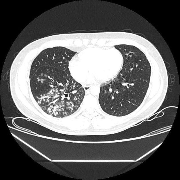
The tree-in-budsign is a nonspecific imaging finding that implies impaction within bronchioles the smallest airway passages in the lung.
. The differential for this finding includes malignant and inflammatory etiologies either infectious or sterile. Multiple causes for tree-in-bud TIB opacities have been reported. Frequency and significance on thin section CT.
Methods cases with tib opacities in the radiology report in 2010 were. Tree-in-bud TIB opacities are a common imaging finding on thoracic CT scan. Gross et al found distal airways disease in all lung specimens and bronchiolar dilatation in 36 of specimens71 HRCT demonstrates bronchiectasis or bronchial wall thickening in 2035 and centrilobular tree-in-bud opacities57 61 67Increased susceptibility to infection may be the underlying cause of bronchiectasis67.
The tree-in-bud pattern is commonly seen at thin-section computed tomography CT of the lungs. Although initially described in 1993 as a thin-section chest CT finding in active tuberculosis TIB opacities. Originally and still often thought to be specific to endobronchial Tb the sign is actually non-specific and is the manifestation of pus mucus fluid or other.
Tree In Bud Opacities Radiology. The purpose of this study was to determine the relative frequency of causes of TIB opacities and identify patterns of disease associated with TIB opacities. Crossref Medline Google Scholar.
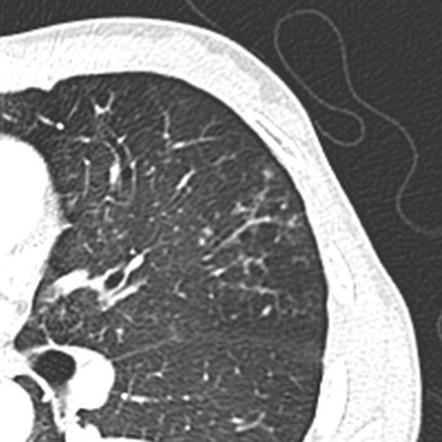
It consists of small centrilobular nodules of soft-tissue attenuation connected to multiple branching linear structures of similar caliber that originate from a. Identification and evaluation of centrilobular opacities on high-resolution CT. In radiology the tree-in-bud sign is a finding on a CT scan that indicates some degree of airway obstruction.
The tree-in-bud sign is a nonspecific imaging finding that implies impaction within bronchioles the smallest airway passages in the lung. What does tree-in-bud opacities mean. Cryptogenic organizing pneumonia 119.
Radiology scientific expert review panel. We investigated the pathological basis of the tree-in-bud lesion by reviewing the pathological specimens of bronchograms of normal lungs and contract radiographs of the post-mortem lungs manifesting active pulmonary. Usually somewhat nodular in appearance the tree-in-bud pattern is generally most pronounced in the lung periphery and associated with abnormalities of the larger airways.
Thus the bronchioles resemble a branching or budding tree and are usually somewhat nodular in appearance. Tree in bud opacification refers to a sign on chest CT where small centrilobular nodules and corresponding small branches simulate the appearance of the end of a branch belonging to a tree that is in bud. What does tree-in-bud opacities mean.
Franquet T et al. 3 Gruden JF Webb WR. It was initially used by JG Im to describe the endobronchial spread of Mycobacterium tuberculosis.
1 However since its first use in 1993 the tree-in-bud pattern has been associated with multiple etiologies. Tree in bud opacification refers to a sign on chest ct where small centrilobular nodules and corresponding small branches simulate the appearance of the end of a branch belonging to a tree that is in bud. J Comput Assist Tomogr 1996.
Thrombotic microangiopathy of pulmonary tumors. As its name implies this pattern resembles a budding tree in CT scans see Fig. Tree-in-bud pattern at thin-section CT of the lungs.
Our Radiology Information System was searched for the term tree-in-bud from January 1 2010 to December 31 2010 iden-tifying 599 examinations. Histopathology The tree-in-bud pattern seen on CT represents radiologic sequelae of an infectious or inflammatory process. The tree-in-bud sign is a nonspecific imaging finding that implies impaction within bronchioles the smallest airway passages in the lung.
In radiology the tree-in-bud sign is a finding on a CT scan that indicates some degree of airway obstruction. In radiology the tree-in-budsign is a finding on a CT scan that indicates some degree of airway obstruction. The tree-in-bud-pattern of images on thin-section lung CT is defined by centrilobular branching structures that resemble a budding tree.
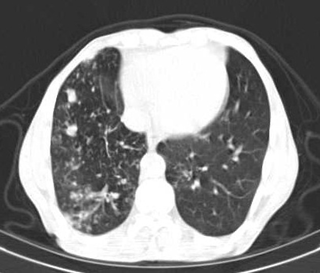
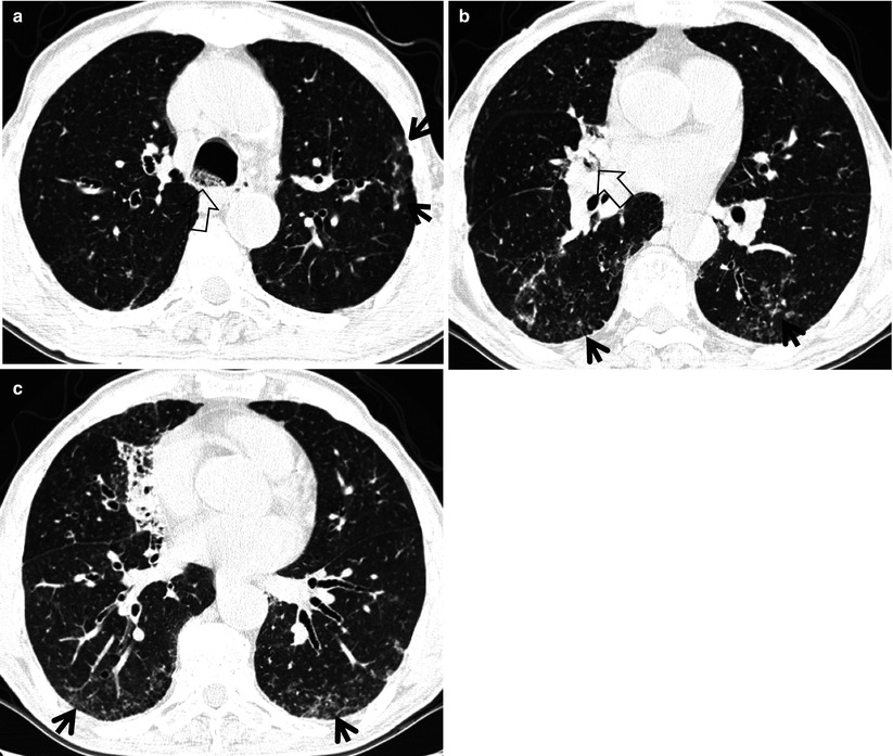
Nodular opacities with tree-in-bud appearance can be associated with other changes in lung parenchyma-such as thickening of the bronchial walls consolidations andor areas of. The tree-in-bud sign is a nonspecific imaging finding that implies impaction within bronchioles the smallest airway passages in the lung. 50 year old male with cough.
TBMAI Tumor emboli rare Chapter 02_32_58_F 22006 236 PM Page 50. These small clustered branching and nodular opacities represent terminal airway mucous impaction with adjacent peribronchiolar inflammation. Centrilobular nodules tree-in-bud pattern ground-glass opacities and consolidation.
2 Aquino SL Gamsu G Webb WR Kee ST. Of these 182 cases were excluded for the following reasons. CT confims numerous centrilobular nodules with opacified distal bronchioles tree-in-bud sign and bronchiectasis.
78 indicating the absenceresolution of TIB opacities 26 incomplete thoracic CT scan studies 75 duplicate. A vascular cause of tree-in-bud pattern on CT. Poorly defined centrilobular nodules and tree-in-bud pattern.
AspirationKartageners Inflammatory plugging PUS. Rossi SE et al. It is usually pronounced in centrilobular branching structures in the lung periphery associated with diseases of the small airways 36The tree-in-bud sign indicates bronchiolar.
The Oral Boards Primer CA2 NODULES MASH POX Metastatic disease Alveolar microlithiasis Silicosissiderosis Histoplasmosis Pox Varicella TREE IN BUD OPACITIES CT MIT Mucous plugging. Tree-in-bud refers to a pattern seen on thin-section chest CT in which centrilobular bronchial dilatation and filling by mucus pus or fluid resembles a budding tree. An unusual pattern similar to the nodules seen in subacute.
Chest x-ray in a 60 year old patient of Asian extraction demonstrates faint reticulonodular opacities. Tree-in-bud pattern seen on high-resolution CT HRCT indicates dilatation of bronchioles and their filling by mucus pus or fluid. Various viral pneumonias 232 especially postbone marrow transplant 173.
Tree in bud opacification refers to a sign on chest ct where small centrilobular nodules and corresponding small branches simulate the appearance of the end of a branch belonging to a tree that is in bud. AJR Am J Roentgenol. However to our knowledge the relative frequencies of the causes have not been evaluated.
These findings most likely represents pulmonary TB or MAC despite negative induced sputum specimens. In radiology the tree-in-bud sign is a finding on a CT scan that indicates some degree of airway obstruction.

Tree In Bud Pattern Radiology Case Radiopaedia Org
2
2
2
Tree In Bud Appearance Endobronchial Spreading Of Pulmonary Tuberculosis Radiology Case Radiopaedia Org
Tree In Bud Sign Lung Radiology Reference Article Radiopaedia Org

Pdf Tree In Bud Semantic Scholar

Tree In Bud Sign Lung Radiology Reference Article Radiopaedia Org
Tree In Bud Sign Lung Radiology Reference Article Radiopaedia Org

Tree In Bud Sign And Bronchiectasis Radiology Case Radiopaedia Org
View Of Tree In Bud The Southwest Respiratory And Critical Care Chronicles
Tree In Bud Sign Radiology Key

Tree In Bud Sign Lung Radiology Reference Article Radiopaedia Org

References In Causes And Imaging Patterns Of Tree In Bud Opacities Chest
2

Tree In Bud Caused By Haemophilus Influenzae Radiology Case Radiopaedia Org
2

Tree In Bud Sign Lung Radiology Reference Article Radiopaedia Org
Tree In Bud Appearance Radiology Case Radiopaedia Org